

- Home
- About Us
- Service
- Application
- Technology
- Resource
- Contact Us

10x Visium HD Spatial Transcriptome
Product Overview
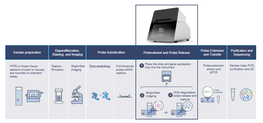
10x Visium HD Spatial Gene Expression Technology represents the cutting edge of high-resolution spatial transcriptomics. It delivers whole-transcriptome data at single-cell resolution, leveraging a novel slide design and a workflow supported by Visium CytAssist to generate high-quality data from human or mouse tissue samples. Since its launch, Visium HD has demonstrated powerful discovery capabilities. The platform is now compatible with both formalin-fixed paraffin-embedded (FFPE) and fresh frozen (FF) samples, enabling flexible sample selection tailored to research needs and empowering researchers to gain in-depth insights into the complexity and functionality of biological samples.
Advantages
 Multi-platform Spatial Omics Solutions
70+ Tumor Tissue Dissociation Expertise
Faster, Cost-saving & Stringently Controlled Library Prep & Sequencing
Multi-omics Integrated Analysis Solutions
Product Parameters
| Indicator | Parameter |
|---|---|
| Sample Types | Fresh frozen/FFPE |
| Sample Format | FFPE blocks/slices OCT blocks |
| Species Types | Human/Mouse |
| Resolution | 2 μm |
| Detection Area | 6.5 mm × 6.5 mm |
| Sequencing Platform | NovaSeq X Plus PE150 |
| Sequencing Data Volume | FFPE: 100 Gb FF: 210 Gb |
| TAT (with/without BI) | 55–65 WD/35–45 WD |
Sample Requirements
| Sample Type | Sample Volume per Sample | Storage Conditions | Transport Conditions |
|---|---|---|---|
| FFPE Block | 1 block | Sealed, stored at 4°C | Transport with ice pack |
| FFPE Section | Quality control sample: Paraffin roll with total thickness ≥50 μm (100 μm for puncture samples) Formal sample: 2–4 adjacent sections, section thickness 4–6 μm |
Quality control sample: Sealed dry in 1.5 mL EP tube, stored at 4°C Formal sample: Newly cut sections mounted on manufacturer-recommended adhesive slides (see sample requirements for specific model); sealed dry, stored at 4°C for ≤30 days (interval between sectioning and library construction) |
|
| OCT Embedded Block | Sample thickness >1 mm, area <6.5 × 6.5 mm | Stored in -80°C freezer; mark the cutting surface on the embedding box | Transport with dry ice |
Workflow
Analysis Contents
| Analysis Type | Analysis Content |
|---|---|
| Standard Analysis |
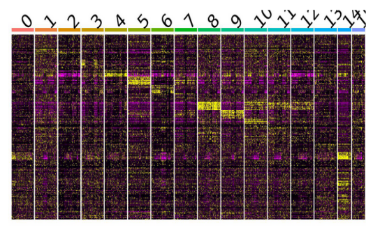
1. Sequencing data statistics and quality control 2. Data quantification 3. Spot filtering 4. Spot subpopulation analysis 5. Cluster highly expressed gene analysis 6. Functional enrichment analysis of cluster highly expressed genes |
| Advanced Analysis |
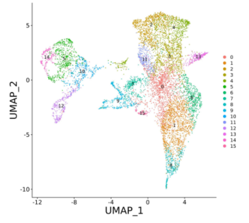
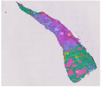
1. Copy number variation analysis 2. Proportion prediction and analysis of malignant vs. normal cells 3. Spatial cell-cell interaction analysis 4. Spatial cell type deconvolution analysis 5. Spatial cell optimized communication analysis 6. Spatial clonal evolution analysis 7. Spatial pseudotime trajectory analysis 8. Gene set scoring and comparison analysis |
Application Scenarios
Oncology: Tumor microenvironment, heterogeneity, progression, morphology & tertiary lymphoid structures
Immunology: Immune cell infiltration, gene expression signatures & dissemination
Developmental Biology: Spatial gene expression in human development & morphology-related gene identification
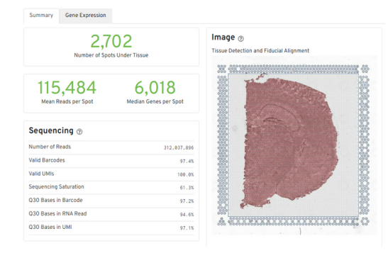
Neuroscience: Brain cell layer mapping & expression profiling of normal/pathological regions
Pathology: Gene-expression enhanced morphological conclusions & pathological accuracy verification
FAQ
Q: What are the advantages of spatial transcriptome applications?
A: Spatial transcriptome sequencing can simultaneously capture gene expression information and spatial distribution data in tissues. It enables the acquisition of transcriptome data from cells at different positions within a single intact tissue section. With broad application prospects in biomedical research, disease mechanism studies, and drug development, spatial transcriptomics helps to deeply understand cellular functions and interactions.
Q: What are the sample quality inspection requirements?
A: To ensure sample quality and reduce experimental risks, we will cut 10–20 tissue sections (10 μm thick) for total RNA extraction and quality testing. It is recommended to proceed with subsequent experiments only for embedded tissue samples with an RNA Integrity Number (RIN) greater than 7.